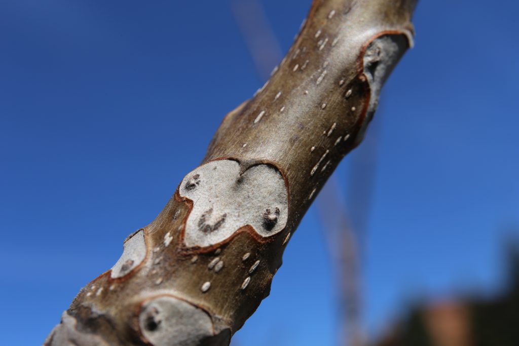
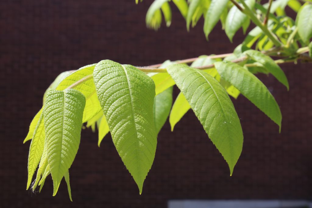
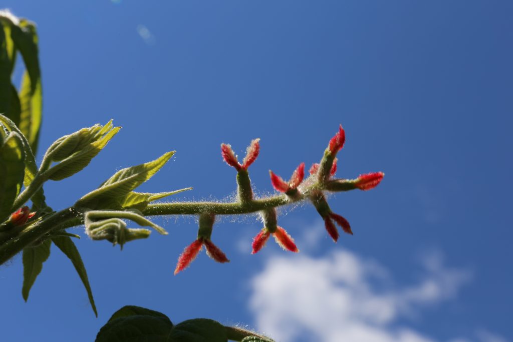
Before reaching your "lichen" goal, note the young tree on your left, tucked against the nursing building. This is a specimen of butternut Juglans cinerea (Fig 12-1, and a-b) with its characteristic large leaf scars, exposing the dark spots where vascular tissue (xylem and phloem) connected to the previous season's leaf. Figures 12-1a and b show young leaf and female flower (which will produce the nut if fertilized).
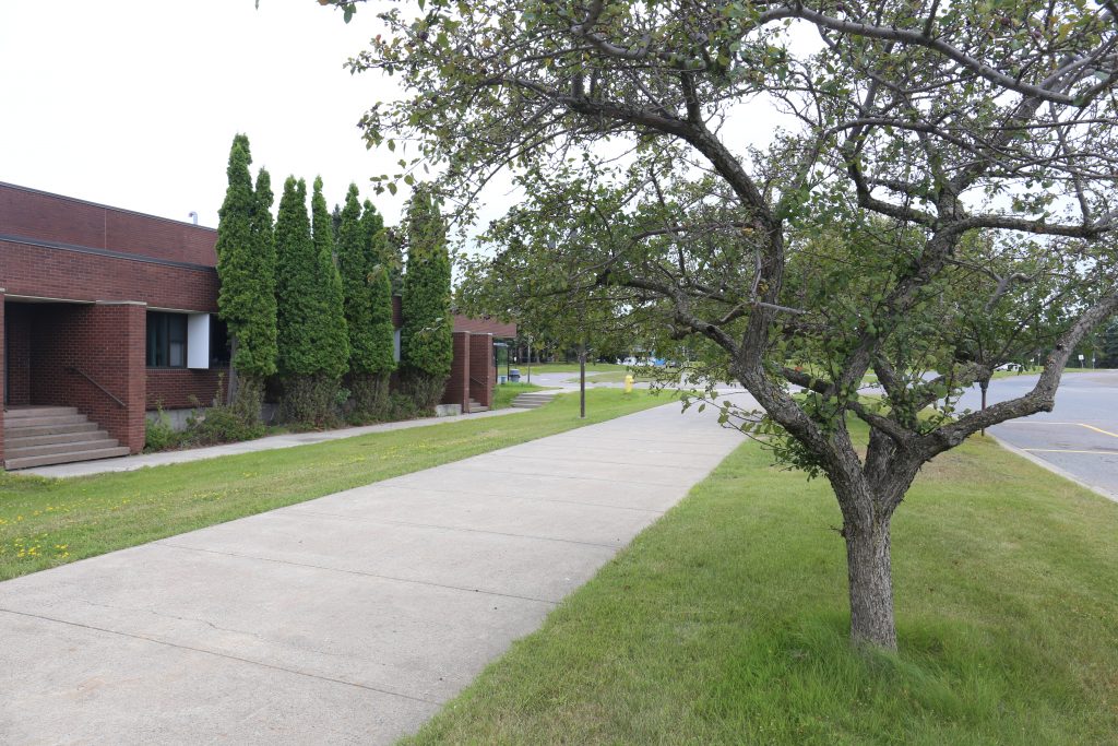
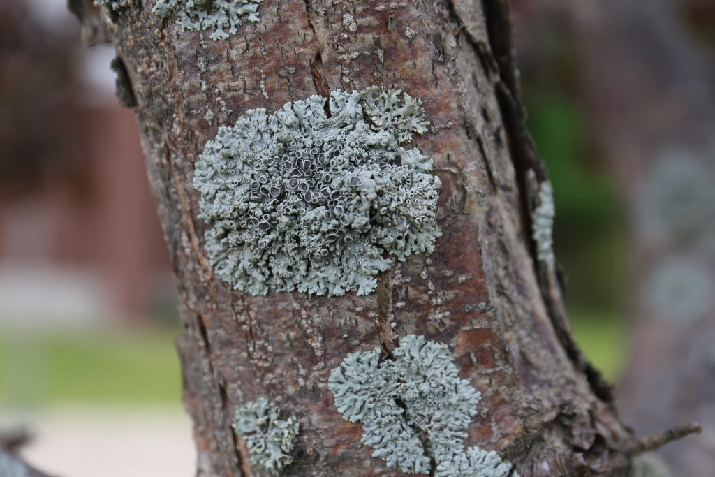
Walking back the way you came between the ATAC and Ryan Buildings, pass along the NE side of the medical school and nursing buildings (Fig. 12-2). Here is a row of a few crab apple trees, again covered with lichens.
The most prominent lichen here are large colonies of hooded rosette lichen (Physcia sp.). Note the incurved ends of the lobes, nearly forming tubes, and the prominent black apothecial disks.
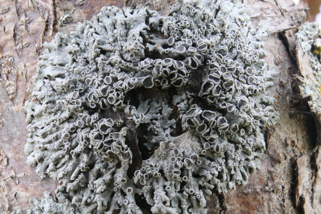
Several species of hooded rosette lichen (Physcia) occur in our region, and they can be difficult to differentiate. P. stellaris seems to be the "simplest" of all, smallish colonies, no cilia, no white pruina or waxy maculae on the surface and no spore producing soredia or any chemical reaction. Common P. aipolia has white pattern of maculae and its internal medulla reacts positively with KOH. P. phaea is similar (also KOH positive) but only occurs on rock and lacks the greyish frosting on surface of apothecia. Physcia adscendens has characteristically long cilia, and P. millegrana is covered with prominent soralia, especially at the tips of lobes. P. caesia seems to be very similar to P. phaea, except it has no apothecia, but rather powdery bluish grey soralia, also favouring rock and occasionally wood (but not bark).